About
The Biosensors for bioengineering group is a junior group under IBEC’s Tenure Track scheme.
Our research is focused on multi tissues organs-on-a-chip (OOC) and more specifically in the metabolic crosstalk within tissues and their relationship with metabolic diseases. Our projects are focused on four key tissues regulating glucose homeostasis, namely, the pancreas, liver, skeletal muscle, and adipose tissue. To achieve this objective, it is necessary a combined interdisciplinary approach.
Biomaterials and tissue engineering research
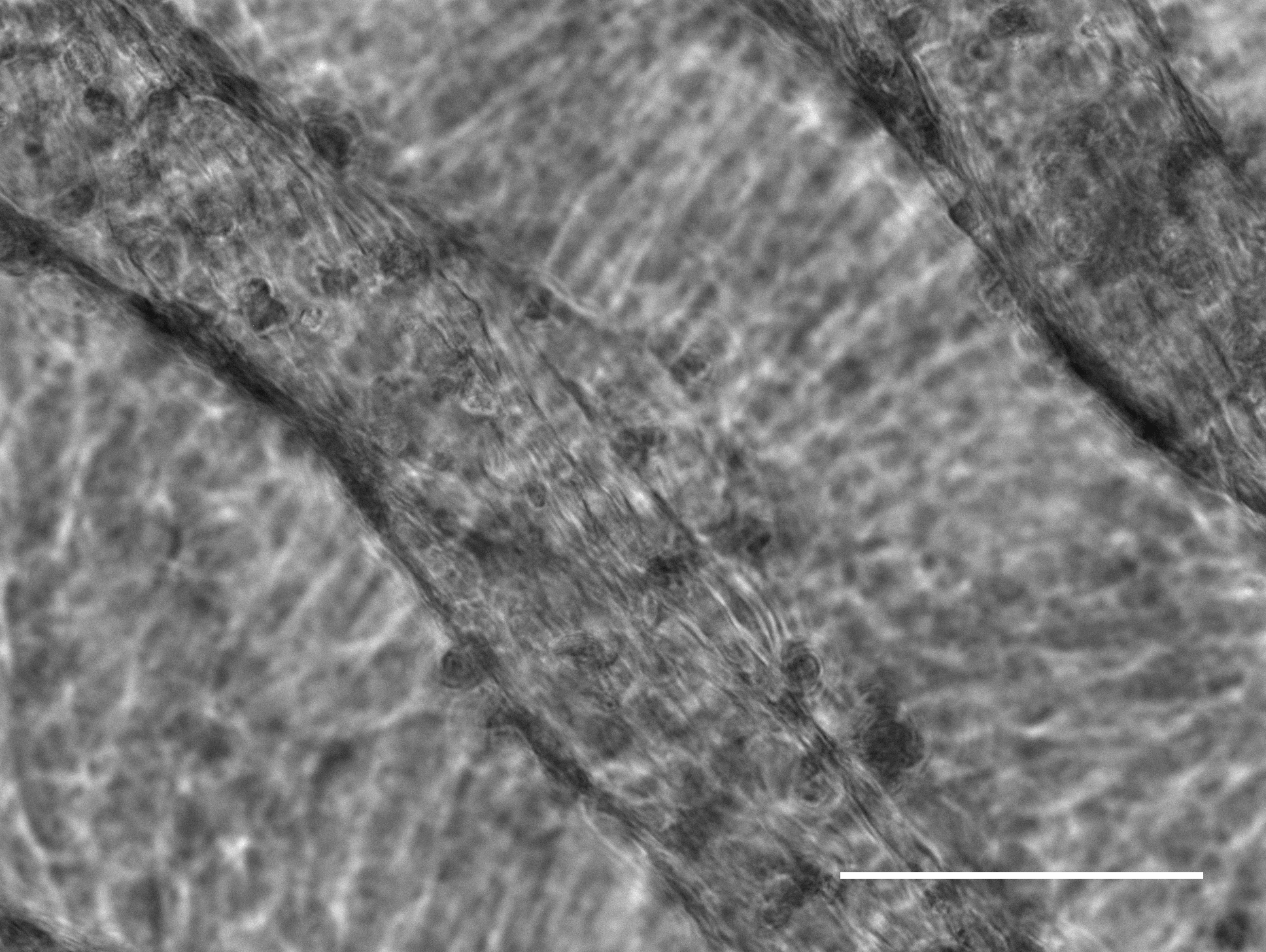
1) We have several lines of research related with skeletal muscle. Our first approach was with C2C12 mice cell line. We evaluated the influence of mechanical stiffness and geometrical confinement on the 3D culture of myoblast-laden chemically modified gelatin photo-cross linkable composite hydrogels in terms of in vitro myogenesis.
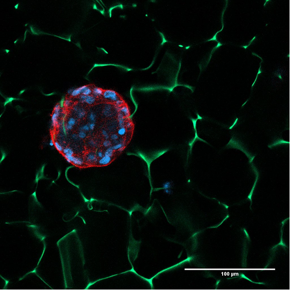
2) Encapsulation of beta-cells like from human skin fibroblast (collaboration with IDIBAPS). This work addresses two critical issues in the design of an efficient beta-cell replacement therapy: an accessible cell source for generation of substitute beta-cells and an adequate delivery device for transplantation. On one hand, we propose to generate transplantable functional insulin-producing beta-cells from fibroblasts through direct reprogramming strategies that bypass the pluripotent iPS stage. On a second objective, we are working in a new system of encapsulating beta-cells like in two steps, microencapsulation to protect cells from immune system and microencapsulation to mechanically protect them and manipulate them.
3) We are developing three-dimensional micro liver models using various biomaterials to recreate the in vivo-like mechanical properties and using hepatocytes and stellate cells. We are collaborating with Grifols company to test some drugs in our model.
4) We have a collaboration project with NovoNordisk to work in new approaches to encapsulate retinal cells.
Biosensing technology:
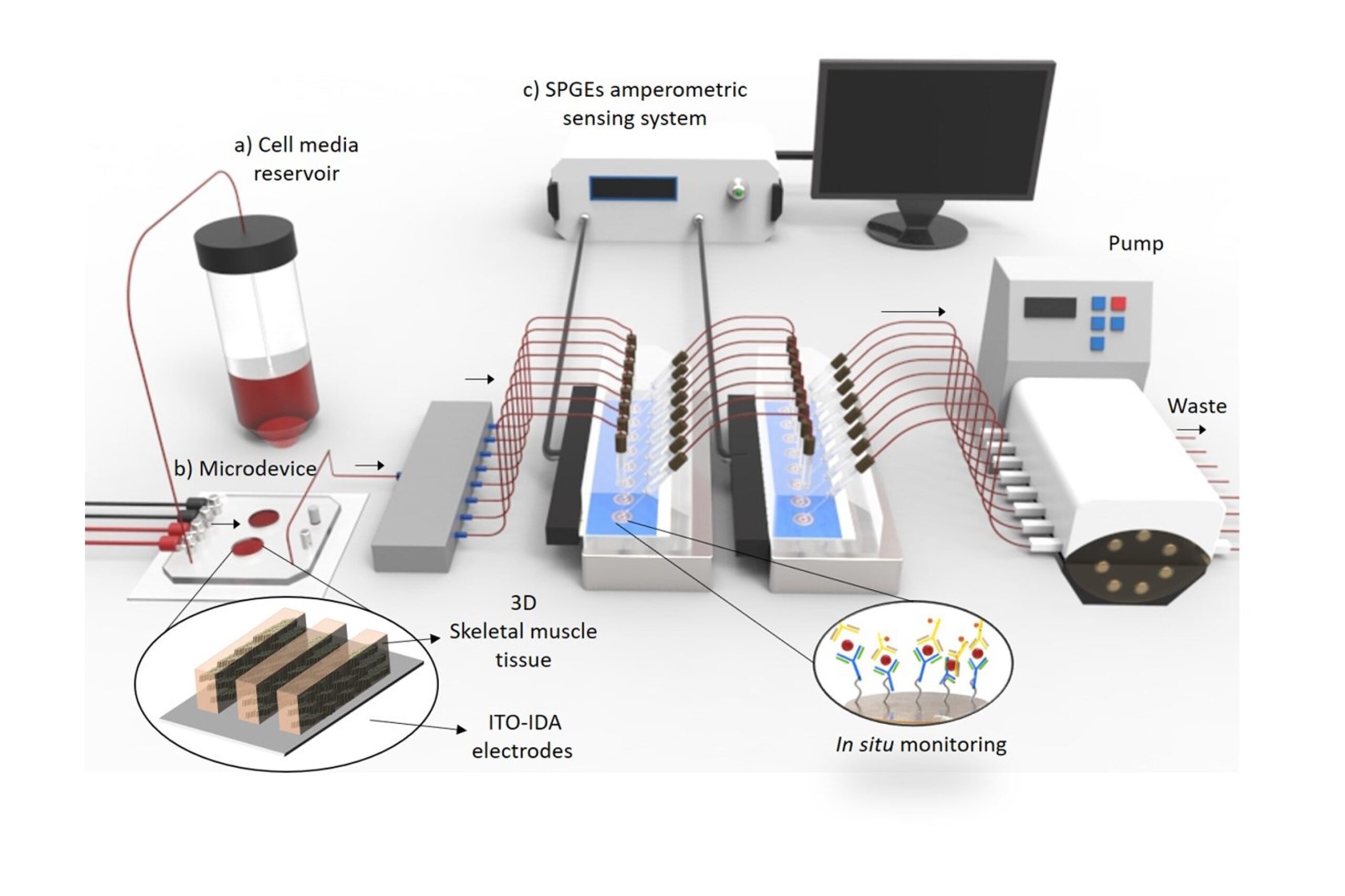
1) Integrating biosensors in an organ-on-a-chip. We are studying with in situ electrochemical biosensors the release of insulin under the effect of external stimuli, changes in glucose levels and myokines secreted by skeletal muscle (multi-OOC approach).
2) Related with this project we are implementing new biosensors systems. To fully exploit the potential of the organs-on-a-chip, there is a need to interface them to integrated sensing modules, capable to monitor in real-time their biochemical response to external stimuli, like stress or drugs. The goal of this project is to answer this need, by developing a novel technology based on integrating localized surface plasmon resonance (LSPR) sensing module to organs-on-a-chip devices to monitor disease and evaluate drug response in organs-on-a-chip models.
3) Myotonic dystrophy type 1 (DM1) (collaboration with Hospital de la Fe and INCLIVA, Valencia, Spain). We have developed human skeletal muscle micro physiological tissues using micro molding technology and we have integrated them with amperometric biosensors to study the inflammatory process related with electrical and chemical stimuli. We have used transdifferentiated skin fibroblast human cells from DM1 patients and healthy human. Using this platform, we have started to evaluate different treatments, to screen drugs and to evaluate doses.
4) NMR integrated with OOC. The objective of this project is to develop a new technology based on magnetic resonance spectroscopy and imaging using dynamic nuclear polarisation (DNP-MR) integrated with OOC devices to monitor disease and evaluate drug response in OOC models. As a proof-of-concept, this project will fabricate a biomimetic multi OOC integrated device composed of liver spheroids and pancreatic islets and develop the necessary DNP-MR hardware and software to study metabolic diseases and for future drug screening applications. We are working in collaboration with Oxford instrument and Multiwave companies.
Staff
Javier Ramón Azcón
 ibecbarcelona.eu
Projects
| NATIONAL PROJECTS | FINANCER | PI |
|---|---|---|
| Development of a “Muscle-on-a-Chip” (MoC) platform for the preclinical evaluation of potential therapies for Duchenne muscular dystrophy (2020-2022) | DUCHENNE ESPAÑA, IV Convocatoria Ayudas a Proyectos de Investigación | Juanma Fernandez |
| BLAD · BioLiver Assist Device (2020-2021) | AGAUR, Ajuts per a projectes innovadors amb potencial d’incorporació al sector productiu – LLAVOR | Javier Ramón |
| INNOTEC- Javier Ramon- Naturfiltr (2021-2023) | TECNIO | Javier Ramón |
| ASITOC Atomic-Sensor-Integrated Tissue-On-a-Chip: optically detected biomagnetism to understand muscular diseases (2021-2022) | BIST_Barcelona Institute of Science and Technology | Juanma Fernandez |
| INTERNATIONAL PROJECTS | FINANCER | PI |
|---|---|---|
| DAMOC · ‘Diabetes Approach by Multi-Organ-on-a-Chip’ (2017-2022) | ERC | Javier Ramón |
| BLOC · Benchtop NMR for Lab-on-Chip (2020-2022) | European Comission FET-Open | Javier Ramón |
| PRIVATELY FUNDED PROJECTS | FINANCER | PI |
|---|---|---|
| Tatami · Therapeutic targeting of MBNL microRNAs as innovative treatments for myotonic dystrophy (2019-2022) | Fundació bancaria “La Caixa” | Javier Ramón |
| FINISHED PROJECTS | FINANCER | PI |
|---|---|---|
| Programa Faster Future 2020: COVID-19 (2021) | Fundraising | Javier Ramón |
| INDUCT · Fabrication of a biomimetic in vitro model of the intestinal tube muscle wall: smooth muscle-on-a-chip (2018-2020) | MINECO | Javier Ramón |
Publications
(See full publication list in ORCID)
[br]
Equipment
Micro and nanofabrication techniques:
- 3D microstructures on hydrogel materials
- Mini-bioreactor for 3D cell culture
- Microelectrodes fabrication
- Synthesis and chemical modification of polymers and surfaces
- Dielectrophoretic cells and micro particles manipulation
Characterization techniques:
- Optical Microscopes (white light/epifluorescence)
- Electrochemical techniques (Potentiometric/Amperometric/Impedance spectroscopy)
- Immunosensing techniques (Fluorescence ELISA/Colorimetric ELISA/magneto ELISA)
Equipment:
- Microfluidic systems (High precision syringe pumps/Peristaltic pumps/Micro valves)
- Biological safety cabinet (class II)
- Epifluorescence microscope for live-cell imaging
- Pulsar – a high-resolution, 60MHz benchtop NMR spectrometer from Oxford Instruments
Access to the Nanotechnology Platform (IBEC Core Facilities): equipment for hot embossing lithography, polymer processing and photolithography, chemical wet etching, e-beam evaporation and surface characterization (TOF-SIMS)
Access to the Scientific and Technological Centers (University of Barcelona): equipment for surface analysis (XPS, AFM, XRD), organic structures characterization (NMR) and microscopy techniques (SEM, TEM, confocal)
Collaborations
- Prof. Josep Samitier
IBEC - Dr. Elena Martinez
IBEC - Dr. Anna Novials
Institut d’Investigacions Biomediques August Pi i Sunyer (IDIBAPS) - Dr. Ramon Gomís
Institut d’Investigacions Biomediques August Pi i Sunyer (IDIBAPS) - Dr. Eduard Montanya
The Bellvitge Biomedical Research Institute (IDIBELL) - Prof. Enric Bertran
Physics and Engineering of Amorphous Materials and Nanostructures (FEMAN), Department of Applied Physics, University of Barcelona - Dr. Montserrat Costa
2020, Director Plasma Proteins Research, Bioscience Industrial Group, Grifols, Barcelona Spain, Collaborative project - Tryfon Antonakakis
2019, Co-Founder & CEO Multiwave Technologies AG 3 Chemin du Pré Fleuri 1228, Geneva Switzerland, FET-open project - Robert Hardy
2019, Project Manager Oxford Instruments plc Abingdon, Oxfordshire, England, FET-open project - Dr. Carlos Villaescusa
2018, Principal Scientist/Specialist, Project Leader, Department of Stem Cell Discovery, Novo Nordisk Denmark, Collaborative project
Clinical collaborations
- Project “TATAMI” funded by Fundación Bancaria “La Caixa” – CaixaHealth program. In this project, we are developing a platform to perform drug screening analysis in human engineered microtissues in close collaboration with Professor Ruben Artero from Instituto de Investigaciones Clínicas de Valencia (INCLIVA) and medical doctor Vilchez from Hospital de la Fe (Valencia)
- We are also collaborating with Hospital de Sant Pau (Barcelona), with the group of senior professor Isabel Illa Sendra we are developing human microtissues to study the myasthenia gravis neuromuscular rare disease.
- In a Smart Specialization Project (RIS3CAT, ADVANCECAT project), I am working with senior professor Eduard Montanya from Hospital de Bellvitge (Barcelona) to develop transplantable patches of human pancreatic islets.
- Finally, we are collaborating with Doctor Jesus Castro from Hospital de la Vall de Hebron (Barcelona) to study chronic fatigue.
News
L’IBEC desenvoluparà òrgans en un xip en tres projectes Pathfinder
BuonMarrow, OMICSENS i PHOENIX-OoC són els tres projectes en els quals el Grup de Biosensors per a la Bioenginyeria de l’IBEC desplegarà el seu ampli coneixement en l’àmbit de biosensors i òrgans en xip. Els projectes, que es desenvoluparan amb el finançament del prestigiós programa Pathfinder Open del Consell d’Innovació Europeu, prometen millorar els tractaments oncològics i impulsar la innovació en diagnòstics.
Un múscul artificial per estudiar la distròfia muscular de Duchenne
El sistema, desenvolupat per l’IBEC, està fabricat a partir de cèl·lules de pacients i és el primer model 3D de múscul capaç de reproduir el dany que provoca la distròfia muscular de Duchenne. El següent pas serà fabricar una plataforma d’ òrgan-en-un-xip que permeti dur a terme estudis preclínics de fàrmacs contra la malaltia i monitorar el dany muscular de manera més eficient. El treball ha rebut finançament de Duchenne Parent Project Espanya, una associació sense ànim de lucre dirigida per les famílies de nens afectats per aquest tipus de distròfia.
Finançament europeu per al tractament de la diabetis tipus 1 mitjançant bioimpressió 3D
L’investigador de l’IBEC Javier Ramón Azcón ha rebut una “ERC Proof of Concept Grant”. Es tracta d’ un prestigiós finançament que concedeix el Consell Europeu de Recerca per explorar el potencial comercial i social de projectes de recerca duts a terme en institucions europees. El projecte de Ramon, Uniink, s’enfoca en el tractament de la diabetis tipus 1 amb teràpia cel·lular i bioimpressió 3D.
L’investigador de l’IBEC James Eills assistirà a una trobada amb premis Nobel
El Dr. James Eills, investigador de l’IBEC, ha estat seleccionat per assistir al prestigiós Lindau Nobel Laureate Meeting que reuneix destacats joves científics d’arreu del món amb premis Nobel. Enguany, … Read more
Cimera científica per lluitar contra les malalties neuromusculars
Científics i pacients es reuneixen a l’IBEC per buscar noves estratègies de tractament per a aquestes patologies minoritàries El diari Ara va entrevistar a Juanma Fernández Costa, investigador postdoctoral del … Read more
Creen un “gimnàs en un xip” que permetrà estudiar la diabetis i desenvolupar nous fàrmacs per al seu tractament
Coincidint amb el dia mundial de la Diabetis, investigadors de l’IBEC fan públic un estudi en què combinen cèl·lules musculars i de pàncrees en un mateix xip i demostren que … Read more
Bioenginyeria per tractar la diabetis als mitjans
Investigadors de l’Institut de Bioenginyeria de Catalunya (IBEC) liderats pel Professor de Recerca ICREA Javier Ramón, apareixen als mitjans per un recent estudi en col·laboració amb investigadors de l’IDIBAPS, on han desenvolupat petites esferes capaces de respondre a variacions en els nivells de glucosa i produir insulina in vitro.
Innovadores esferes fabricades amb bioenginyeria podrien ajudar contra la diabetis
Investigadors de l’IBEC, en col·laboració amb l’IDIBAPS a Barcelona, han desenvolupat petites esferes capaces de respondre a variacions als nivells de glucosa, i de produir insulina, in vitro. Aquests esferoides biomimètics, i no tòxics, contenen cèl·lules β pancreàtiques i es preparen utilitzant tecnologia de bioimpressió 3D. Aquest enfocament podria ajudar, en el futur, a millorar els resultats clínics de les estratègies de trasplantament de cèl·lules β que es fan servir el tractament de la diabetis, així com per a les plataformes in vitro de desenvolupament de fàrmacs.
Les teràpies regeneratives del futur van confluir al Simposi IBEC, amb experts internacionals i més de 300 inscrits
Més de tres-cents experts internacionals en el camp de la investigació en teràpies regeneratives es van citar al simposi organitzat per l’Institut de Bioenginyeria de Catalunya (IBEC) per presentar els últims avenços en miniòrgans, òrgans-en-un-xip, bioimpressió 3D i enginyeria de teixits, entre d’altres.
Nou biosensor amb alta sensibilitat detecta marcador inflamatori al múscul
En una publicació recent a la revista Nanophotonics, investigadors de l’IBEC presenten un nou biosensor per a la detecció directa i sensible de la proteïna interleucina-6 al múscul, un indicador d’inflamació i de potencial malaltia, demostrant l’alt rendiment del dispositiu en músculs esquelètics obtinguts per bioenginyeria 3D. Aquest nou enfocament pot resultar en una eina prometedora per mesurar l’eficàcia de fàrmacs davant de malalties en què hi ha inflamació, com la distròfia muscular.
Jobs
Postdoctoral Researcher at the Biosensors for Bioengineering Research Group
Ref: PR_JR/Deadline: 09/12/2023
Postdoctoral position at the Biosensors for Bioengineering Research Group
Ref: PD_JR/ Deadline: 10/11/2023
Laboratory Assistant at the Biosensors for Bioengineering Research Group
Ref: LA_JR/Deadline: 22/11/2023
Laboratory Technician at the Biosensors for Bioengineering Research Group
Ref: LT_JR//Deadline: 28th April 2023